XFEL: Crystal structures determined from powders of polymers
Crystal structures determined from powders of polymers
Using a technique tailored to the study of small molecules at the LCLS and European XFEL X-ray lasers, an international team of researchers has resolved the structure of metal–organic polymers that are notoriously difficult to grow into large crystals. The results settle long-standing controversies regarding the atomic connectivity and layer stacking of these compounds.

The FXE instrument at European XFEL
Metal–organic chalcogenolates are self-assembling polymers of a metal and an organic molecule containing sulfur, selenium, or tellurium. The materials have attracted interest for their semiconducting properties, light−matter interactions, and catalytic activity. These compounds suffer from extremely small crystals, which makes it difficult to determine their structure.
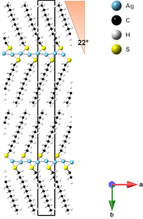
A team of scientists, including researchers from European XFEL, has now used a technique called small-molecule serial femtosecond crystallography (smSFX) at the Linac Coherent Light Source (LCLS) in California and the European XFEL to study microcrystalline silver n-alkanethiolates. After analysis with new computational methods developed specifically for the purpose, the measurements yielded high-quality crystal structures directly from powder suspensions of the compounds.
X-ray free-electron lasers (XFELs) produce a high-flux beam of X-ray pulses with ultrashort duration that makes these facilities ideal for analysing microcrystals at atomic resolution using smSFX. “The smSFX technique relies on the concept of diffraction before destruction to collect diffraction images of samples at room temperature,” explains Christopher Milne, leading scientist at the FXE instrument at which the European XFEL measurements were carried out. “The X-ray pulse duration is shorter than the progression of radiation-induced ionization damage, and the high photon flux of each pulse can produce a strong diffraction signal even from microcrystals.” This capability is particularly useful to study systems that have only small or defective crystals. The SFX technique has been routinely used to investigate protein crystals at XFELs, but has only recently been extended to the study of small molecules.
“We determined the crystal structures of silver n-alkanethiolates with various alkyl chain lengths, over which there had been considerable disagreement,” says James Nathan Hohman from the University of Connecticut, USA, who led the investigation. “These are the first atomic-resolution smSFX data sets. We selected these materials to resolve a few puzzles about their atomic-scale organization. The method is robust, and we showed we could resolve unknown crystal structures in just a few minutes of instrument time. The detail we obtained demonstrates the power of the technique for collecting high-quality structural data on the complex hybrid materials relevant for semiconducting, superconducting, and quantum computing applications.”
The study was carried out by a team led by the University of Connecticut and Lawrence Berkeley National Laboratory, USA, and including researchers from European XFEL, LCLS, and MAX IV Laboratory in Sweden. The results were published in the Journal of the American Chemical Society.
Reference:
“XFEL Microcrystallography of Self-Assembling Silver n‐Alkanethiolates”, Mariya Aleksich, Daniel W. Paley, Elyse A. Schriber et al., J. Am. Chem. Soc. 145, 31, 17042–17055 (2023), DOI:10.1021/jacs.3c0218
Contact:
Dr Christopher J. Milne
Tel: +49-40-8998-5610
E-mail: christopher.milne@xfel.eu
Dr Bernd Ebeling
Tel: +49-40-8998-6921
E-mail: bernd.ebeling@xfel.eu
