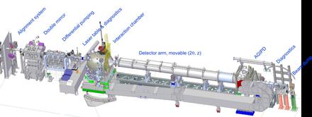
The main instrumentation is installed in two hutches in the experimental hall, the X-ray optics and the experimental hutch. Furthermore, the main optical laser hutch and the Instrument laser hutch are in immediate vicinity to the experimental hutch. The experiments are supervised and monitored from the control room, which includes all necessary computing resources to control all motors and devices in the experiment hutch, optics hutch, and tunnel. Furthermore, the control room is furnished with several working places for instrument operators and for users to perform online data analysis during the experiments.
Instrument specifications

| Bandwidth | 6×10⁻⁵, 10-4, or 10-3 (pink) |
|---|---|
| Photon energy range | 5–25 keV (coherent) and > 25 keV (high-energy option) |
| Bunch charge | 1–1000 pC |
| Polarization | Linear (horizontal) |
| Pulse duration | 1–100 fs |
| Beam size | 1–200 µm, 1 mm, and nanofocus option |
| Special optics |
2 monochromators (Si111 and Si220) 2 compound refractive lens (CRL) transfocator units Split and delay line High-energy Laue monochromator (optional) Mirror in experiment hutch (for grazing incidence liquid scattering) |
| Equipment |
Multipurpose chamber SAXS/WAXS geometries with long horizontal detector arm Small vertical WAXS setup Single-pulse X-ray diagnostics Different detector systems (AGIPD, FastCCD) Optical pump laser source |
Source and Optics
X-ray beam energies and pulse patterns
-
energy tunable in a range from 5 to 25 keV
-
pink SASE bandwith about 10-3
-
two additional monochromators available Si-111, ΔE/E = 1.4 × 10⁻⁴; Si-220, ΔE/E = 5.9 × 10⁻⁵
-
-
Self-seeding will be implemented first at MID/SASE2 (not available for EUE)
-
pulse energy ~0.5 mJ = 3 1011 photons/pulse at 10 keV
-
pulse duration <100fs
-
10 pulse trains/s
-
Intra-bunch up to 4.5 MHz
-
i.e. Δt = 220 ns spacing between single pulses
-
Integers nΔt possible (n=4 tested)
-
1, 30, 120, ..., 2700 pulses/train
-
Focussing
Split and Delay line (not available for EUE)
A split-delay line (SDL) will be available to create pairs of photon pulses out of a single pulse. The pulse pairs will arrive at the sample with a delay from 0 to 800 ps with a precision down to a few fs. The SDL operates in a range from 5 to 10 keV and furthermore allows the two pulses to be either collinear or inclined with respect to each other. Like this both pulses can hit the sample but due to the inclination the scattering images will be separated at the detector. Schemes where the X-ray pulse pairs are combined with an optical pump laser pulse can be realized and in this manner ultrafast structural dynamics can be spatially encoded into the scattering images.
fs optical laser source
All beamlines at the European XFEL will have an optical laser that is capable of matching the pulse schemes of the European XFEL. The laser radiation, generated in the hutch of the Optical Lasers group, will have a pulse length between 100 fs and sub-15 fs, a wavelength of 800 nm, and power up to 3 mJ per pulse. To enable optical laser radiation tailored to different requirements, the laser beam will be guided into an instrument laser hutch. Here, various optical parameters, e.g. intensity, energy, beam size, and pulse length, can be modified depending on the experimental needs. The laser hutch is located next to the MID instrument, hence providing the shortest delivery pathway to the benefit of stability and dispersion. In case of extremely dispersion-sensitive beam parameters, the optical laser beam can also be optimized directly in front of the experiment chamber. Here, the timing diagnostics is located to determine the temporal jitter between the arrival of the optical laser and the XFEL beam.
Experimental hutch

Multi-purpose sample chamber
The experiment chamber is located ~959.5 m after the source point. It will be operated under moderate vacuum conditions of ~10−5–10−1 mbar. In order to garantee windowless operation of the whole beamline, the chamber will be connected via a differential pumping section to the UHV part of the beamline (~10−9 mbar). If the samples or sample environment do not tolerate vacuum, the experiment chamber can be separated from the beamline upstream using a transparent diamond window.
It is possible to install various sample environments such as liquid jet injectors or a fast sample exchanger for solid samples. Setups to heat, cool, or apply a magnetic field to the sample will be available as well. Samples can also be mounted on a hexapod table with the necessary degrees of freedom for scanning and alignment purposes. A second hexapod will be available to host lenses for short working distance nano focussing. Additionally, the chamber will feature beam diagnostics and alignment tools and allow coupling-in the optical laser beam.
Detector configurations
Several different detector configurations can be achieved at the MID instrument. The option to operate a very long (8 m) horizontal scattering arm is a special feature of the instrument. The horizontal arm can move continuously in an angular range from 0° to 50°. For highest position precision of the detector, a high quality floor is installed in the MID hutch, on which the long detector arm can move via pressurized air pads. Furthermore, the sample to detector distance can be changed by driving the detector cage along the long detector arm.
Detector: AGIPD 1M
The AGIPD 1M is the main pixel detector of MID, with the capability of resolving pulses inside a train (4.5 MHz). The pixel size is 200 micron and the detector can be located up to 8 m downstream. It can also be placed as close as 20 cm, or less if the sample environment is adapted, to the sample. In forward scattering geometry a central hole in the detector allows the direct beam to pass without any damage to the sensor. Via a vacuum interface on the back side of AGIPD and a beam pipe, the direct, transmitted beam is sent down towards an end-station where the spectrum, intensity and beam position is monitored before the beam is blocked and fully absorbed by a beam stopper.
| Parameter | Parameter value | Comment |
|---|---|---|
| Energy range | QE > 80% in the range 3.5–13 keV | 22% QE at 25 keV |
| Dynamic range | 0–34 Me-pulse-1pixel-1 | > 104 ph at 12.4 keV, 4x10⁴ ph at 3 keV, 14-bit counter |
| Frame rate | 4.5 MHz | — |
| Noice (ENC) | About 359 e-rms (100 eV) | Before irradiation, values after irradiation under investigation |
| Storage cells | 352 | Max. images per train |
| Sensor thickness | 500 µm | Silicon sensor |
| Pixel size | 200 µm x 200 µm | — |
| Number of pixels (total) | 1024 x 1024 (physical) | Double-sized pixels between ASICs on the same module |
| Central beam hole | Yes | Variable hole |
| Mechanical gap between sensor modules | About 3.4 mm (long side) and about 0.4 mm (short side) | — |
Other detectors for MID
| AGIPD 1M | JUNGFRAU 1M | ePix 100 1M | GOTTHARD (-II) | |
|---|---|---|---|---|
| Energy range (keV) | 3-25 | 3-25 | 3-20 | 3-25 |
| Dynamic range | 104 ph/px/pulse @ 12keV | 104 ph/px/pulse @ 12keV | 100 ph/px @ 8keV | 104 ph/px/pulse @ 12keV |
| Pixel size | 200 × 200 µm2 | 75 × 75 µm2 | 50 × 50 µm2 | 50 (25) µm |
| Noise | ~1000eV | ~200eV (HG) | <200eV | <750eV |
| Repetition rate | 4.5 MHz | Currently 200kHz | 120Hz | 800kHz (4.5 MHz) |
| Number of storage cells | 352 | 16 | - | (Compact storage for full pulse train) |
| In-vacuum | Yes | Yes | Yes | No |
| (#mod) Array size | (4) 110×110 mm2 /mod | (2) 40×80 mm2 /mod
|
(2) 35×38 mm2 /mod. | (1) ~6×64 mm2 1280 (2560) pxl |
Diagnostic Endstation
The diagnostic end-station of the MID instrument offers beam characteristics such as spectral distribution, intensity and position on a shot-to-shot basis. The spectral characterization is achieved by the use of a bent crystal spectrometer, using both Si and diamond crystals. For detection, a Gotthard I line detector with 50mm strip size (later upgrade to Gotthard II 25mm pixel size) is used. In two places downstream of the spectrometer, the intensity and position can be characterized using diamond detectors. Additionally an imager with a medium and high resolution option is available to characterize the beam shape on a lower temporal scale.
-
Download
Technical Design Report: Scientific Instrument MID