XFEL: New simulations enable faster biological insights
New simulations enable faster biological insights
Single-particle X-ray diffraction imaging (SPI) is a technique used by European XFEL’s SPB/SFX instrument to look at biological molecules in their native environments. By accurately simulating the SPB/SFX instrument’s detector, researchers have determined a route that could result in increased data-collection efficiency, as well as an improvement in the resolution of the images taken through SPI. This could enable new insights into the structure and evolution of biological molecules.
Detectors can blur, distort or obscure the signals they are attempting to detect, making it harder for scientists to understand collected data. The researchers used the simulations to replicate the noise in their detector, carrying out an investigation to show the optimal number of image snapshots they would need to take of a biological protein to accurately render its structure.
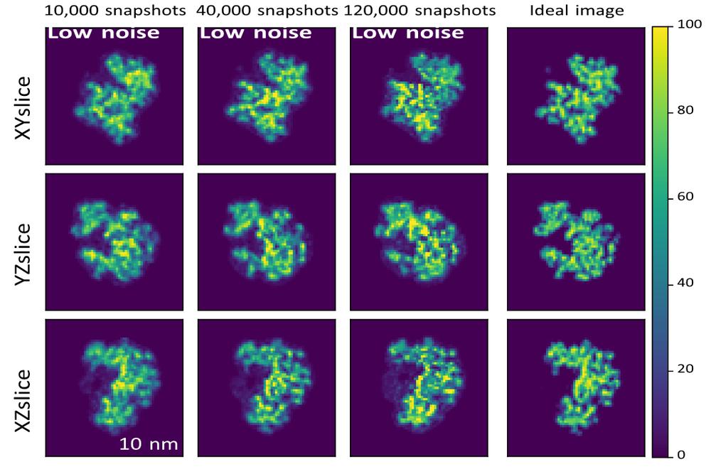
Simulations of the structure of biological molecules for different numbers of image snapshots as compared with the ‘perfect’ image, shown right. After a certain number of snapshots, the improvement in the image becomes incremental, or insignificant, allowing an ‘optimum’ number of snapshots to be determined. [Juncheng E, Yoonhee Kim and Chan Kim, European XFEL].
The results will improve the efficiency with which we can image biological samples in their native environments, as well as ever increasing imaging resolution.
Read more:
Juncheng, E., et al. "Expected resolution limits of x-ray free-electron laser single-particle imaging for realistic source and detector properties." Structural Dynamics 9.6 (2022)
Available here.
Scientific contact:
Richard Bean
Tel: +49-40-8998-6843
E-mail: richard.bean@xfel.eu
Press contact:
Bernd Ebeling
Tel: +49-40-8998-6921
E-mail: bernd.ebeling@xfel.eu
