XFEL: Researchers snap giant virus in 3-D
Researchers snap giant virus in 3-D
Using the world´s most powerful X-ray laser, an international team of researchers, including scientists from DESY and European XFEL, have produced a 3-D image of an intact, infectious virus. The experiment at the SLAC National Accelerator Laboratory in the U.S. reveals part of the inner structure of the Mimivirus, as the collaborative team led by Dr. Tomas Ekeberg from Uppsala University in Sweden report in the journal Physical Review Letters.
The study using SLAC´s Linac Coherent Light Source (LCLS) demonstrates how the 3-D structure of many types of biological samples can be reconstructed from a series of X-ray laser snapshots. This technique holds great potential, also for the European XFEL, an X-ray Free-Electron Laser that is currently under construction between DESY in Hamburg and the neighbouring town of Schenefeld.
“Ever since I started in this field of X-ray laser research, this has always been the dream – to acquire 3-D images of real biological samples,” said Dr. Tomas Ekeberg, a biophysicist at Uppsala University in Sweden and lead author of the study. “This is fantastic – it´s a breakthrough in our research.”
The Mimivirus belongs to a curious class of giant viruses discovered just over a decade ago. With a diameter of 750 nanometers, including a hairy fur, Mimivirus is larger than some bacteria and it was misclassified as a bacterium until 2003. Subsequent discoveries have found other giant viruses, some of which are even larger. Mimivirus is also genetically complex, with nearly 1000 major genes compared to only a handful in the Aids virus HIV.
Scientists have been trying to determine the inner structure of these giant viruses to learn more about their origins: For example, did they borrow genes over time from the host organisms they infect, like amoebas? Did they precede cell-based life or devolve from cell-based organisms?
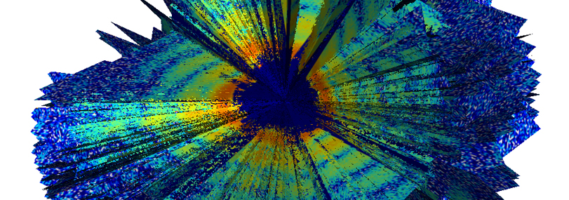
Each image captured a projection of a separate virus particle at a random orientation, so the collection of images of viruses in different orientations provided a more complete, 3-D view. “Our study provides just a glimpse of the potential of future investigations of three-dimensional biological structures with X-ray lasers like the LCLS or the European XFEL”, said co-author Prof. Henry Chapman, DESY scientist from the Center for Free-Electron Laser Science CFEL in Hamburg.
"The paper provides a proof of concept", said co-author Janos Hajdu, a professor at Uppsala University and an advisor to European XFEL. "We show that the problem of orientation recovery can be solved and thus 'getting rid of the crystal' in X-ray diffraction experiments is possible."
While the technique used at LCLS did not provide high-resolution details of the internal virus structure in this demonstration study, it did confirm that its contents are lopsided, with an area that appears more densely concentrated. “We can see quite clearly that the inside of these viruses is not uniform,” Ekeberg said. This same general feature had also been seen before using an electron-based imaging technique with frozen samples. X-ray lasers like the LCLS and the future European XFEL allow studies of viruses and other biological samples in a more natural, intact state. According to the researchers, this technique shows promise for achieving sharper images that reveal more inner details in the future because of the uniquely intense, penetrating power of the X-rays.

