Capabilities of the FXE instrument

Current capabilities of the FXE instrument (state in April 2024)

FXE instrument started user operation in September 2017 [A. Galler , et al., J. Synch. Rad., 26, 1432 (2019)]. Several components and standard setups have been commissioned and validated during recents years by performing measurements, including pump-probe experiments, or developing new methodologies to perform new research. On this page we present current experimental parameters of the instrument and its standard components with their performance specifications. Additional components in the commissioning phase and under development are given in the section 4. 

This page is being constantly updated and currently still in construction. To receive most recent information on specific topics please contact the FXE-support mailing list (fxe-support@xfel.eu) or directly the FXE Leading Scientist Chris Milne (christopher.milne@xfel.eu).

1. Main experimental parameters currently reachable at FXE

Typical experimental parameters currently available for experiments at the FXE instrument are listed below (state on November 2020), but subject to changes without specific notice.

Parameter
Value
Unit
Photon energy (range)
4.9 – 20
keV
Pulse number/train
single shot – 300
---
Intra-train rep.-rate
1128, 564, 376, etc.
kHz
Pulse energy (at source)
1 - 4
mJ
Bandwidth (SASE)
ca. 2*10-3 (SASE) and 10-4 (Si mono)
dE/E
Pointing stability
~ 2
urad
X-ray focus size
~ 8 - 200
um
Timing jitter (fwhm)
70
fs

2.  Standard setup for pump-probe spectroscopy and scattering in solution jets 

The setup is optimized for performing femtosecond optical pump/X-ray probe experiments mainly in solutions of molecular complexes or suspensions of nanoparticles. Optical excitation is typically done using frequency harmonics and fundamental wavelength of femtosecond radiation at 800 nm (typ. 50 fs duration fwhm) and 1030 nm (typ. 800 fs duration fwhm), wavelength tunable source is also available for the 50 fs pulses (section 2.4). X-ray probing of photo-induced transient dynamics in solution is currently limited to X-ray emission spectroscopy performed by 16-crystal von Hamos spectrometer (2.2) and wide-angle X-ray scattering using the Large Pixel Detector (2.3). For more details also see [D. Khakhulin, et al., Appl. Sci., 10, 995 (2020)] and [F. Lima et al. J. Synch. Rad. 30, 1168 (2023)].

Picture
Arrangement of main components around the interaction region
Picture
Setup for simultaneous X-ray emission and X-ray scattering, top view

2.1 Sample environment

The main sample environment is designed as a sealed chamber with Kapton windows large enough to allow simultaneous collection of X-ray emission and wide-angle X-ray scattering. The sample chamber can be filled with an inert gas (typ. He or N2) to ensure anaerobic or dry atmosphere. The chamber is equipped with SMA feedthroughs for installing additional 0D detectors. There are three view ports for sample microscopes and one window for the laser excitation beam to enter the chamber at an angle of 10-15 deg with respect to the X-ray beam. The main X-ray beam propagates in a He-purged flight tube and enters the chamber directly without a window. 

Solution samples are circulated in an HPLC pump based system and supplied through quartz nozzles with round opening of various diameters, typ. 100 um, 50 um or 150 um. With a 100 um round nozzle the typical linear speed of a water jet reaches ca. 60 m/s allowing to refresh the sample volume between individual pump-probe events at the repetition rate of up to 564 kHz.

Picture
Standard sample environment for liquid chemistry experiments

Additionally to the main pump two sample circulation pumps are available for in-line static UV-VIS spectroscopy and solution/solvent refill, all pumps are controlled remotely. The nozzles can be manipulated by three XYZ translation stages, a YAG fluorescence screen and fast diode can be mounted next to the nozzle and brought into the beam without opening the chamber.

2.2. von Hamos X-ray emission spectrometer 

Wavelength dispersive von Hamos spectrometer is based on an array of 16 independently controlled cylindrical analyzer crystals with 0.5 m bending radius and allows measuring entire emission lineshapes in a single X-ray shot (or train) without scanning. Emission spectra from individual crystals are focused and overlapped on a Jungfrau 500k or 1M detector running at 10 Hz framerate, i.e. one image per X-ray train. Due to the flexibility and large number of analyzers simultaneous collection of multiple emission lines from the same or different elements can be performed, provided a compatible configuration of analyzer crystals exists and feasible. The setup geometry accommodates Bragg angles in the range 67-83 degrees. Details on how this approach has been scaled towards working at MHz repetition rates can be found in [M. Biednov et al. Nucl. Inst. and Meth. A 1055, 168540 (2023)].

Below we provide an example of simultaneous full Ni K-line emission spectrum collected from a 10 mM aqueous solution of a Ni salt flowed in a 100 um round jet. Incoming X-ray photons at 9.3 keV with 150 pulses per train were used for this measurement.

Picture
Single-train Jungfrau image for a nickel complex in aqueous solution (C = 10 mM) 100 µm jet, incoming photon energy 9.3 keV, 0.564MHz, 150 pulses/train, 3 mJ/pulse
Picture
Ni emission lineshapes extracted from the Jungfrau images shown above after averaging of ca. 20 min of data collection

2.3. Large Pixel Detector (LPD) for wide-angle X-ray scattering 

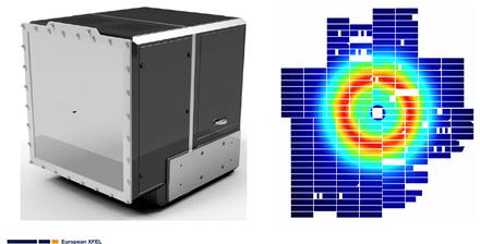
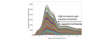
Wide-angle X-ray scattering on solutions systems is collected using LPD at a typical detector distance of about 20-30 cm. The LPD-1M is a modular detector consisting of 16 super-modules, 256x256 pixels each, with 500 um square pixels and 500 um thick silicon sensor. LPD offers 3 gain stages with respective gain reduction factors of ca. 8.7 and 107 for the medium and low gain stages respectively, thus enabling simultaneous dynamic range of ca. 104 at 12 keV. X-ray scattering images can be recorded in the 510 total memory cells at the repetition rate of up to 4.5 MHz thus storing up to 510 single-shot X-ray images per pulse train. The unique feature of LPD is the parallel gain readout technology so that the final image is constructed from individual pixels each in the optimal gain stage by software-defined thresholds excluding possible analog gain switching uncertainties. That means that now when the gain transitions have been characterized and thresholds optimized the detector response is expected to be linear over the entire dynamic range covered by the three gain stages.

Picture
Left: The Large Pixel Detector (LPD). Right: Typical scattering pattern on LPD from a 100 um water jet
Picture
Signals of various intensities in a single pixels in the parallel (left) and automatic (right) gain readout modes.
Picture
Scattering curves produced by azimuthal integration of single-shot LPD images from 100 um water jet for X-ray shots of different intensities

2.4. Sample excitation capabilities 

Currently only optical radiation is available for excitation at FXE. Laser radiation from the main Pump-Probe Laser system of European XFEL with 800 nm and 1030 nm wavelength is readily available together with their frequency harmonics (see table below). The wavelength-tunable source, TOPAS,  has been installed and commissioned in the hutch and is planned to be used in a pump-probe experiment starting 2021 (see the measured tuning curve below).

Picture
TOPAS tuning curve measured at FXE

The optical excitation beams are delivered to the sample interaction region using a flexible optical setup able to accommodate various configurations in terms of excitation geometry and wavelengths (see images below). This includes collinear and non-collinear (15 degrees) laser  incoupling, ability to change excitation wavelength within one shift preserving the focusing conditions (currently not including TOPAS).

Picture
Optical setup for sample excitation with various optical wavelengths

3. Standard setup for single-shot X-ray diffraction in transmission geometry

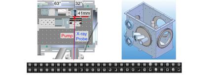
The setup consisted of a vacuum chamber (down to 10-5 mbar) with Kapton window for forward scattering typically collected by the LPD (section 2.3).  The optical pump and the X-ray probe beams are focused on the sample at normal incidence in quasi-collinear geometry. The direct X-ray beam is off-centered on downstream Kapton window and on the detector behind to enable large Q-range. Thin film samples for single-shot measurements are commonly mounted on Si3N4 membranes in a standard frame with typical pitch of 0.5-1 mm between the membranes. Fast XY translation stages allow to center membrane targets on the X-ray beam using predefined optimal positions currently at a rate of ca. 0.5 Hz. Once the target has reached the position a single pair of X-ray and optical laser pulses is delivered upon a trigger from the control system. 

Picture
Left, Arrangement of interacting beams and sample mounting stage inside the vacuum chamber. Right, vacuum chamber for diffraction experiments in transmission geometry. Bottom, sample frame.

4. Components in the commissioning phase or under development 

The component and capabilities described below are currently under commissioning at FXE and their availability is yet limited. Should you be interested in using this equipment please inquire with the instrument staff.

4.1. Setup for simultaneous grazing incidence XRD and emission spectroscopy on solid state systems

At FXE we have performed initial tests of the grazing incidence diffraction setup using a Z-axis goniometer in vertical geometry (see photograph below). Additionally the von Hamos spectrometer was aligned to collect X-ray emission from the surface of the sample and two Jungfrau detectors were used respectively.

Picture
Combined pump-probe GI-XRD and XES setup at FXE

4.2. X-ray absorption spectroscopy by simultaneous scanning of the undulator gap and cryogenic Si (111) monochromator.

Femtosecond X-ray absorption capability of the FXE instrument is currently under commissioning. X-ray energy scanning using a 2-bounce monochromator geometry has been demonstrated on standard metal foils (Fe, Co, Cu and Au) in transmission geometry and mM concentration liquid solutions using total fluorescence yield. Since the undulator gap of the XFEL source can be scanned as well, it is possible to perform spectroscopic measurements in an extended energy range (EXAFS).

If scanning is not necessary and only a monochromatic beam at fixed energy is needed for an experiment, it can be readily provided during user operation. Due to beam headload issues the number of X-ray pulses per train reliably transmitted through the monochromator is currently limited to typical 10-30 pulse/train depending on the photon energy and pulse intensity.   

The setup is currently not offered for the upcoming 7th call for proposals, however is planned to be fully commissioned in the upcoming run (2021-I) together with users, so may become available by the second half of 2021. Please inquire with the instrument staff.

Picture
Left, Cryogenic 4-bounce monochromator at FXE. Right, typical EXAFS scan in transmission on Fe foil

4.3. Johann-type X-ray emission spectrometer

FXE is equipped with a Johann-type X-ray emission spectrometer consisting of 5 independently controlled spherical analyzer crystals with 1m radius of curvature. The emission signals are collected by a detector on the robotic arm that can be either Jungfrau area detector or a 0D APD depending on the application. Initial commissioning of the spectrometer scatting has been performed and the Co K-beta emission from a foil was acquired on a Jungfrau detector. Further commissioning on simultaneous scanning of the crystals and the detector following the Rowland circle is ongoing.

Picture
5-crystal Johann spectrometer (left) and Co Kb (center) and VTC (right) X-ray emission spectra scanned with one crystal on a foil