XFEL: Unveiling finer details in the physics of materials

https://www.xfel.eu/e35178/e35455/e35456 https://www.xfel.eu/news_and_events/news/index_eng.html news news news eng 1 1 10 both 0 1 %Y/%m/%d Press-Release
eng
2023/02/21
Back

Unveiling finer details in the physics of materials

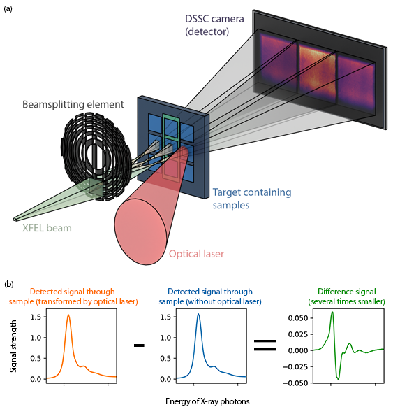
Scientists at the European XFEL’s SCS instrument routinely use a technique called transient X-ray absorption spectroscopy (XAS) to investigate materials that have applications in data storage and processing, catalysis, or in the search for room temperature superconductors. Investigating very small changes in the motion of electrons within a material’s structure on ultrashort timescales provides scientists with fingerprints of the complex processes at play within them. This helps them characterise samples that are important for energy and materials research.

Using the European XFEL’s brilliant pulses, researchers can overcome some of the issues of conventional transient XAS---such as long measurement times---but the varying intensity of European XFEL’s pulses provides its own challenges. Now, scientists at SCS have implemented a new sampling scheme for improving the efficiency of such measurements.

Download [1.8 MB, 5416 x 1968]
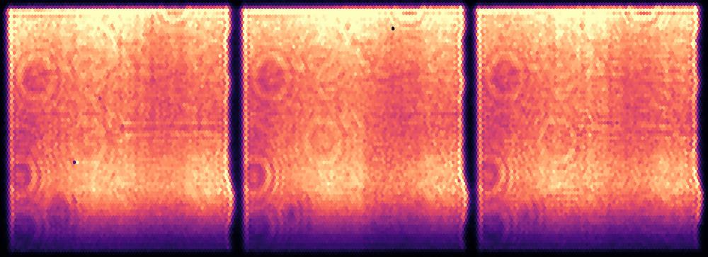
Fig 1: Images of the three detected signals, from the three separate beams generated by the new method described by scientists at SCS.
Fig 2 (a): The X-ray beam is split into three copies. Two of these copies are passed through identical samples of the material under investigation, with one of these samples also being illuminated by a laser ('optical laser' in the figure). This transforms it into a new state, interesting to researchers. From this, scientists are able to 'subtract' detrimental noise, revealing the finest details of the sample under investigation. (b): A simplified cartoon showing how scientists 'subtract' signals in the three-pulse method. In the left-most image (orange) is the signal detected when one of the three X-ray beam copies through the sample transformed into a new state by an optical laser. Next to it in the centre (blue) is a signal from a second copy of the X-ray beam, passed through a piece of the sample that has been left undisturbed. By subtracting the blue from the orange, scientists can obtain the right-hand signal (green) which is several times smaller than the other two. This enables them to see the finest changes in a sample during an experiment.
The scheme, characterised in the Journal of Synchrotron Radiation, involves splitting the pulses from the European XFEL into three copies. One copy measures fluctuation in the incoming X-ray pulses from the XFEL, while the remaining two copies probe two identical samples of the material under investigation, one illuminated by a laser to trigger a reaction. The simultaneous measurements reduce the impact of the XFEL varying on a pulse to pulse basis, and can reveal the finest details of the sample under investigation. This provides a large improvement on the sensitivity of the method compared with traditional synchrotron results.

 “We believe this technique has the potential to improve the efficiency at which XFEL scientists can probe interfaces or dilute systems on an ultrashort time scale,” says Loïc Le Guyader. “This scheme, only available at SCS, will allow us to further push the boundaries of materials and energy science.”

Publication:

Le Guyader, L., Eschenlohr, A., Beye, M., et al. (2023). J. Synchrotron Rad. 30.
http://doi.org/10.1107/S1600577523000619

Scientific contact:

Loïc Le Guyader
Tel: +49-40-8998-6708
E-mail: loic.le.guyader@xfel.eu

Press contact:

Bernd Ebeling
Tel: +49-40-8998-6921 
E-mail: bernd.ebeling@xfel.eu