XFEL: New technique reduces sample volume and waste
New technique reduces sample volume and waste
A research team comprising scientists from Arizona State University, US, and European XFEL has developed a unique technique that can dramatically reduce waste of precious biological samples. This paves the way to structural analysis and molecular movies of substances that are difficult to isolate in larger quantities. The team also revealed new details of a protein which has an important function in Gram-negative bacteria, with potential applications for the development of new antibiotics. The results of this new approach have been published in Nature Communications.
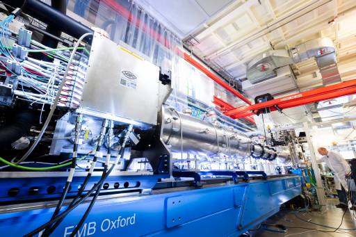
The serial femtosecond crystallography (SFX) instrument at European XFEL gives researchers the possibility to study the intricate details of protein complexes. However, one drawback has been that the commonly used sample delivery method, which continuously jets the protein crystal suspension into the path of the X-ray flashes, wastes more than 99% of the sample due to the pulsed nature of the beam.
The research team, led by Alexandra Ros, professor in Arizona State University's School of Molecular Sciences and the Center for Applied Structural Discovery in the Biodesign Institute, developed a 3D-printed microfluidic device that can deliver a stream of sample-laden droplets segmented with immiscible oil. The researchers then produce the droplet segmentation at the same rate as the X-ray laser pulses, and deliver the sample laden droplets in a fast-moving liquid microjet with the sample ideally present only during exposure to the X-ray pulses, thereby reducing the waste.
The sample studied is an enzyme known as KDO8PS (3-deoxy-D-manno-octulosonate-8-phosphate synthase), which is vital for the survival and virulence of most Gram-negative bacteria. This makes the enzyme a potential target for antibiotic studies. Ros and her collaborators were able to reveal new details in a previously undefined loop region of the enzyme. In addition, they were able to show that the diffraction quality of the KDO8PS crystals was similar both when injected with their new technique using the microfluidic device and by the more conventional continuous injection technique. The use of the microfluidic device, however, resulted in ~60% reduction in sample consumption giving a solid reasoning for future studies at X-ray free electron lasers (XFEL) such as the European XFEL to adopt the microfluidic device that delivers sample-laden droplets segmented with oil.
More Information:
News release by Arizona State University: New microfluidic device minimizes loss of high value samples
