XFEL: Low background noise crucial for single particle imaging experiments
Low background noise crucial for single particle imaging experiments
Taking snapshots of single molecules with X-rays has long been a dream for many scientists. Such experiments have successfully been computationally modelled, but have never been practically demonstrated before.
In a model experiment carried out at the European Synchrotron Radiation Facility (ESRF), European XFEL scientists, together with international collaborators, have now come one step closer to successfully carrying out so-called single particle imaging experiments (SPI) at X-ray laser facilities such as European XFEL. In a paper published today in the journal from the International Union of Crystallography (IUCrJ), scientists demonstrate experimentally that, in principle, a 3D structure can indeed be obtained from many tens of thousands of very weak images, using X-rays with similar properties as produced at X-ray free-electron lasers such as European XFEL.
A single biomolecule, such as a protein, does not scatter X-rays very well and so only produces a very weak signal. In order to compensate for this, single particle imaging experiments require an extremely intense X-ray beam and a highly sensitive detector to produce good quality results. The very high repetition rate of European XFEL now provides a unique opportunity to carry out such experiments. The SPB/SFX instrument at European XFEL has been designed with these issues in mind, and scientists are hopeful that they will be able to use the instrument to carry out these types of experiments with small biomolecules such as proteins in the very near future.
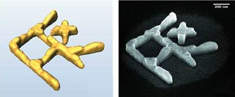
The experiment detailed in the paper published today, was conducted at the ESRF synchrotron and was designed to simulate the conditions of SPI experiments at then future European XFEL instrument SPB/SFX. Synchrotron beams are a lot less intense than XFEL beams, so, to ensure that the amount of signal reaching the detector was equal to that expected at European XFEL the scientists used a sample made of gold. Gold scatters many more photons than a single biomolecule, thereby compensating for the comparably weak synchrotron beam.
The results show that even with weak signals an accurate 3D model of the sample can be calculated. Crucially however, the experiment also shows that a low level of background noise is vital in order to achieve good results.
“This is an important step towards being able to do single particle imaging properly with X-rays” says Adrian Mancuso, European XFEL leading scientist at the SPB/SFX instrument. “Computer models of single particle experiments are not very good at estimating the amount of background noise we can expect in a real experiment, and how that might affect the results. With this experimental simulation we get a feeling for the problems that we will face during experiments at European XFEL, and show practically for which signal-to-background level these experiments can indeed work.”
While the state-of-the-art detectors at European XFEL are able to capture the weak signals produced by single particles, stray photons from other sources will also be picked up, making it harder to differentiate between meaningful signal and background noise. For other types of experiments at XFELs the background noise is negligible in comparison to the amount of photons hitting a sample, so instruments are not usually designed with the possibility to clean up the beam. The SPB/SFX instrument at European XFEL, however, has been designed with exactly this in mind. Over the last year, scientists and engineers at European XFEL have been working step-by-step to optimize the beam with the goal of performing SPI experiments with small biomolecules.
The first SPI experiments conducted at European XFEL, studied relatively large particles such as viruses that scatter more X-ray light. Experiments with increasingly smaller and, therefore, weaker scattering molecules are planned for the near future.
Original article:
Experimental 3D coherent diffractive imaging from photon-sparse random projections
K. Giewekemeyer, A. Aquila, N.-T. D. Loh, Y. Chushkin, K. S. Shanks, J.T. Weiss, M. W. Tate, H. T. Philipp, S. Stern, P. Vagovic, M. Mehrjoo, C. Teo, M. Barthelmess, F. Zontone, C. Chang, R. C. Tiberio, A. Sakdinawat, G. J. Williams, S. M. Gruner and A. P. Mancuso
IUCrJ 6, 2052 (2019)
https://doi.org/10.1107/S2052252519002781
