XFEL: Capturing the strongest X-ray beam on Earth
Capturing the strongest X-ray beam on Earth
The European XFEL beam. Copyright European XFEL
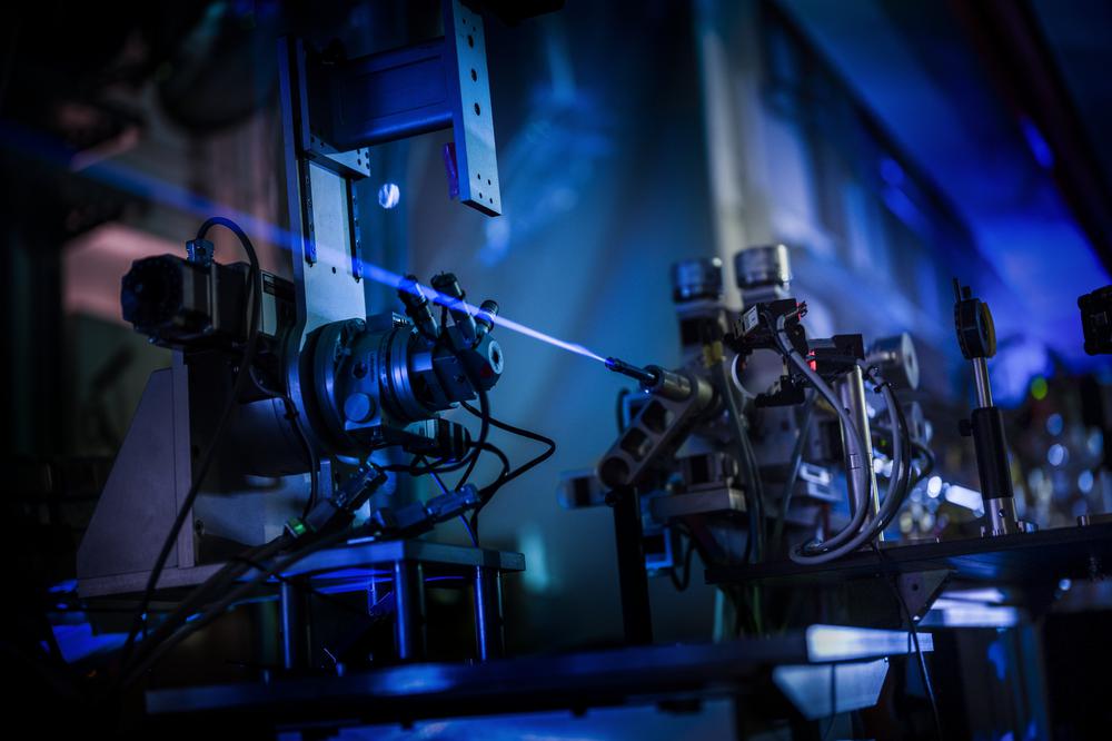
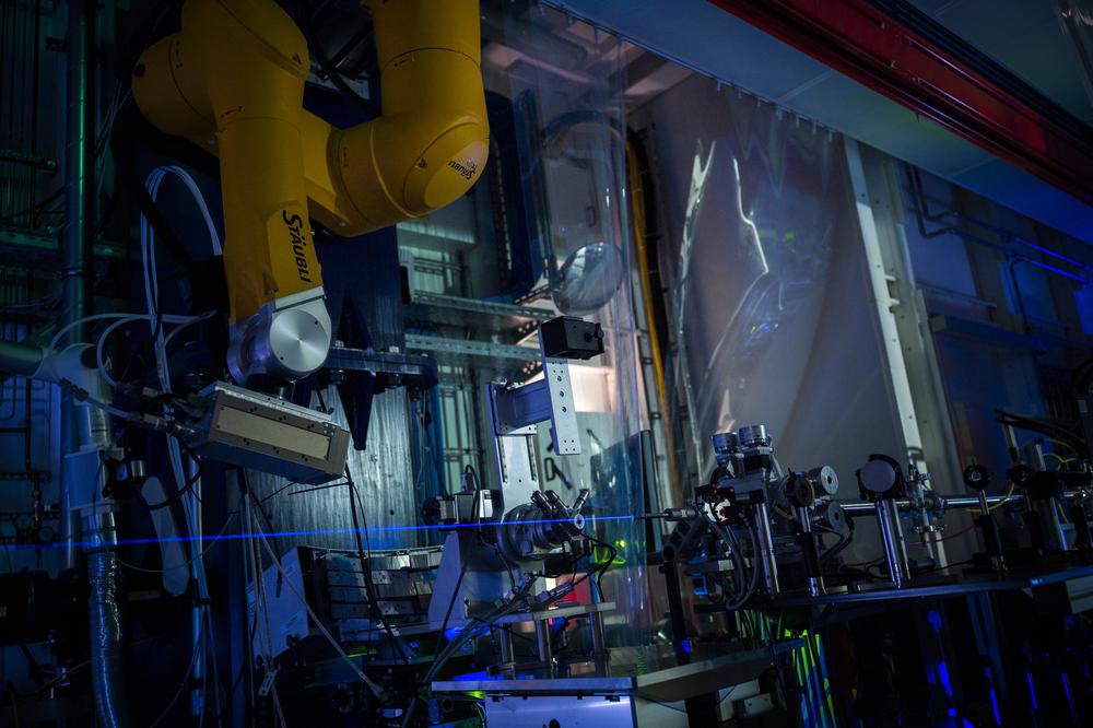
At European XFEL scientists use intense X-rays to take pictures of the smallest particles imaginable. The European XFEL X-ray beam is a billion times brighter than other traditional X-ray sources, but since X-rays are invisible to the naked eye, it is not usually possible to see the X-ray beam. Working together with a professional photographer, scientists at the largest X-ray laser in the world located in Schenefeld near Hamburg, have now managed to capture an image of the intense European XFEL X-ray beam. The pictures were taken as the X-ray beam entered the experiment area in the FXE instrument hutch at the end of a journey that started in a 3.4km long underground tunnel.
On the images published today, the X-ray beam appears as a thin blue stripe. What we are actually seeing, however, is glowing nitrogen molecules which the X-ray beam has caused to light up as it travels through the air thereby interacting with the molecules.
The European XFEL beam in the FXE hutch. Copyright European XFEL / Jan Hosan
"It works much like a fluorescent or neon lamp, where the gas in the centre of the tube lights up when the electric current is turned on”, explains Prof. Christian Bressler, team leader at the FXE (Femtosecond X-ray Experiments) instrument where the new images were made. Even though the European XFEL X-ray beam is extremely intense, the induced glow of the nitrogen molecules is, however, still relatively weak and not so easy to see with the naked eye. Today’s images were only possible when taken in complete darkness and using an exposure time of 90 seconds. The beam as seen in the images comprised 800 pulses per second, and has a thickness of 1mm. While the camera equipment was set-up inside the experiment hutch, the photos were taken remotely from outside the hutch, in the neighbouring control room.
The method used to make the photos does not only lead to pretty results, but also has a scientific use. “With our sensitive detectors, we can use the glowing of the molecules induced by the X-ray beam for monitoring purposes” said Harald Sinn, responsible for X-ray optics at European XFEL.
Images can be found here
See also below the 'making of' video showing how FXE members and photographer Jan Hosan from the agency 'fotogloria' went about getting the images.
