{'subtitle': 'The molecular film captures the very rapid reaction of the enzyme beta-lactamase from tuberculosis bacteria with the cephalosporin antibiotic ceftriaxone in slow motion. ', 'active_start_dt_iso': '09 September 2021 00:00 h CEST', 'active_end_dt_iso': '', 'active_start_dt': '2021/09/09', 'translationofid': 1897, 'keywords': [], 'langs': [u'eng', u'ger'], 'active_end_dt': '', 'title': 'Filming antibiotic resistance in slow-motion', 'media': [{'href': u'media/7873/agipd07.jpg', 'content_type': u'image/jpeg', 'idmedia': 7873, 'filename': u'agipd07.jpg'}, {'href': u'media/7874/agipd07_thumbnail.jpg', 'content_type': u'image/jpeg', 'idmedia': 7874, 'filename': u'agipd07_thumbnail.jpg'}, {'href': u'media/7875/Slide1_thumbnail.JPG', 'content_type': u'image/jpeg', 'idmedia': 7875, 'filename': u'Slide1_thumbnail.JPG'}, {'href': u'media/7876/Slide1.JPG', 'content_type': u'image/jpeg', 'idmedia': 7876, 'filename': u'Slide1.JPG'}], 'active_toggle': 1, 'body': '<p class="newspublisher_hint">Many resistant bacteria produce the enzyme beta-lactamase. This enzyme can render the most commonly used beta-lactam antibiotics ineffective. The team, led by Marius Schmidt of the University of Wisconsin-Milwaukee, have now studied the first step of this inactivation at the SPB/SFX experimental station of the European XFEL, observing how the antibiotic binds to the beta-lactamase from resistant tuberculosis bacteria within milliseconds (thousandths of a second).</p><p class="more">more</p><p><iframe title="YouTube video player" src="https://www.youtube.com/embed/MbUefYBTObw" width="560" height="315" frameborder="0" allowfullscreen="allowfullscreen"></iframe></p><p>Using the X-ray laser European XFEL an international team of researchers has successfully filmed a reaction step that is important for the development of antibiotic resistance. The molecular film captures the very rapid reaction of the enzyme beta-lactamase from tuberculosis bacteria with the cephalosporin antibiotic ceftriaxone in slow motion. The team published the results in IUCrJ, the journal of the <strong><a href="https://journals.iucr.org/m/issues/2021/06/00/mf5055/index.html"><em>International Union of Crystallography</em></a>.</strong></p><p><div class="news-image-container"><img class="news-image" src="media/7873/agipd07.jpg" alt="" width="340" height="233"><div class="news-image-caption">The detector AGIPD at the European XFEL used for the experiment. Copyright: European XFEL</div></div>Antibiotic resistance occurs, in part, when bacteria acquire the ability to inactivate the antibiotics used against them. Many resistant bacteria produce the enzyme beta-lactamase. This enzyme can render the most commonly used beta-lactam antibiotics ineffective. The team, led by Marius Schmidt of the University of Wisconsin-Milwaukee, have now studied the first step of this inactivation at the SPB/SFX experimental station of the European XFEL, observing how the antibiotic binds to the beta-lactamase from resistant tuberculosis bacteria within milliseconds (thousandths of a second). The researchers also investigated how the enzyme inhibitor sulbactam reacts with the bacterial enzyme. Sulbactam, administered at the same time as the antibiotics, binds to the bacterial beta-lactamase, thereby blocking it. As a result the enzyme is unable to inactivate the antibiotics whose effectiveness is restored this way.<div class="news-image-container"><a class="fancybox" href="media/7876/Slide1.JPG"><img class="news-image" src="media/7875/Slide1_thumbnail.JPG" alt="" width="485" height="363"></a><div class="news-image-caption">Experimental setup at the European XFEL. BlaC microcrystals are mixed with substrate and injected into the X-ray interaction region (dotted circle) after a delay determined by the distance between the mixing region and the X-rays, the capillary width and the flow rate. Diffusion of substrate into the crystals occurs during this time. Source: https://journals.iucr.org/m/issues/2021/06/00/mf5055/index.html</div></div>Up to now, crystals have been a requirement to study biomolecules using X-rays, but with the advent of free electron X-ray lasers, researchers are able to study extremely small crystals. In the case of beta-lactamase, the enzyme crystals and the antibiotic solution are mixed together very quickly, and the reaction starts when the antibiotic diffuses into the enzyme crystal. Because of the small size of the crystals, diffusion is very fast, and all the beta-lactamase molecules in the crystal react with the antibiotic solution almost simultaneously. The special mix-and-inject method used in the study requires custom-made injectors, which were provided by the group of Lois Pollack (Cornell University) for use at the European XFEL. The method enables atomically accurate slow-motion imaging of fast biological processes in enzymes and other biomolecules.</p><p>"Enzymes such as beta-lactamase are of great importance for medicine," explains Marius Schmidt. "X-ray lasers like the European XFEL now make it possible to study much smaller crystals than in the past. Our experiment, together with previous results, has shown how X-ray lasers can be used as an important tool for biological research in the future," he adds.</p>', 'change_uid': u'kidambim', 'active': 1, 'dt': '2021/09/09', 'change_dt': '21/09/09 11:19:28', 'topnews': 0, 'lang': 'eng', 'titleimage': u'media/7874/agipd07_thumbnail.jpg', 'idnews': 1897, 'idtitleimage': 7874, 'dtd': '2021/09/09'}eng,ger
2021/09/09
Back
Filming antibiotic resistance in slow-motion
The molecular film captures the very rapid reaction of the enzyme beta-lactamase from tuberculosis bacteria with the cephalosporin antibiotic ceftriaxone in slow motion.
Using the X-ray laser European XFEL an international team of researchers has successfully filmed a reaction step that is important for the development of antibiotic resistance. The molecular film captures the very rapid reaction of the enzyme beta-lactamase from tuberculosis bacteria with the cephalosporin antibiotic ceftriaxone in slow motion. The team published the results in IUCrJ, the journal of the International Union of Crystallography.
The detector AGIPD at the European XFEL used for the experiment. Copyright: European XFEL
Antibiotic resistance occurs, in part, when bacteria acquire the ability to inactivate the antibiotics used against them. Many resistant bacteria produce the enzyme beta-lactamase. This enzyme can render the most commonly used beta-lactam antibiotics ineffective. The team, led by Marius Schmidt of the University of Wisconsin-Milwaukee, have now studied the first step of this inactivation at the SPB/SFX experimental station of the European XFEL, observing how the antibiotic binds to the beta-lactamase from resistant tuberculosis bacteria within milliseconds (thousandths of a second). The researchers also investigated how the enzyme inhibitor sulbactam reacts with the bacterial enzyme. Sulbactam, administered at the same time as the antibiotics, binds to the bacterial beta-lactamase, thereby blocking it. As a result the enzyme is unable to inactivate the antibiotics whose effectiveness is restored this way.
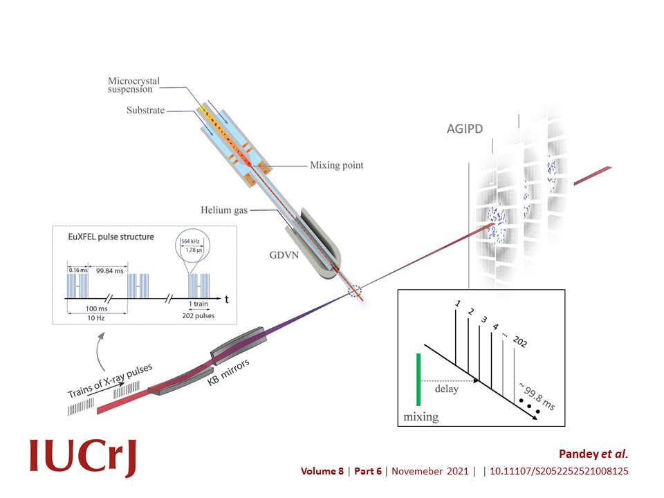
Experimental setup at the European XFEL. BlaC microcrystals are mixed with substrate and injected into the X-ray interaction region (dotted circle) after a delay determined by the distance between the mixing region and the X-rays, the capillary width and the flow rate. Diffusion of substrate into the crystals occurs during this time. Source: https://journals.iucr.org/m/issues/2021/06/00/mf5055/index.html
Up to now, crystals have been a requirement to study biomolecules using X-rays, but with the advent of free electron X-ray lasers, researchers are able to study extremely small crystals. In the case of beta-lactamase, the enzyme crystals and the antibiotic solution are mixed together very quickly, and the reaction starts when the antibiotic diffuses into the enzyme crystal. Because of the small size of the crystals, diffusion is very fast, and all the beta-lactamase molecules in the crystal react with the antibiotic solution almost simultaneously. The special mix-and-inject method used in the study requires custom-made injectors, which were provided by the group of Lois Pollack (Cornell University) for use at the European XFEL. The method enables atomically accurate slow-motion imaging of fast biological processes in enzymes and other biomolecules.
"Enzymes such as beta-lactamase are of great importance for medicine," explains Marius Schmidt. "X-ray lasers like the European XFEL now make it possible to study much smaller crystals than in the past. Our experiment, together with previous results, has shown how X-ray lasers can be used as an important tool for biological research in the future," he adds.