XFEL: Snapshots of exploding oxygen
Snapshots of exploding oxygen

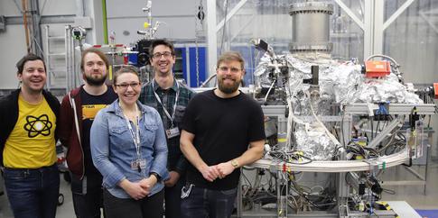
Some members of the scientific team at the SQS instrument at European XFEL. Photo: Markus Schöffler, Uni Frankfurt.
Scientists have long dreamt of being able to see the precise movements in single molecules, such as changes in structure and the forming and breaking of individual bonds. An experimental technique capable of capturing these miniscule movements that happen on extremely fast timescales would increase our understanding of how such processes evolve in detail, and may ultimately give scientists the tools to steer the behavior of ensembles of molecules.

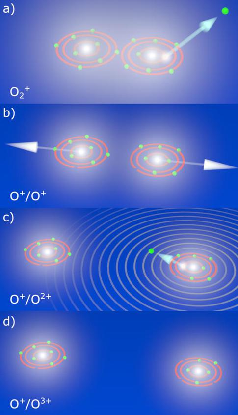
Scheme of photoelectron diffraction imaging during Coulomb explosion of O2. (a) A K-shell electron is ionized upon irradiation with XFEL light. (b) After the innershell vacancy has been filled after a first Auger decay, the molecule is doubly charged and fragments in a Coulomb explosion. (c) During the fragmentation a second photon triggers the emission of another K-shell electron illuminating the molecule from within. (d) A further, subsequent Auger decay yields the observed final O+/O3+ state in which the Coulomb explosion continues until the ions are well separated and finally detected. Steps (a) to (c) occur during a single XFEL light pulse, i.e., within approximately 25 fs. Copyright: Kastirke et al
Over the years, different methods have been proposed to take snapshots of molecules. While X-ray diffraction, for example, is a relatively well-established method for studying nano-sized particles, observing small molecules in the gas-phase that consist of only a few atoms is still very challenging. The movement of the individual atoms occurs on such short timescales that extremely bright and extremely short light flashes are needed to capture the precise details. Such conditions are now accessible at the SQS soft X-ray instrument at European XFEL that started user operation in November 2018. For the first time, experiments can take advantage of the high repetition rates, unique to European XFEL, to carry out detailed studies on individual molecules.
In a paper published today in the journal Physical Review X, a team of scientists led by Till Jahnke from the Goethe University Frankfurt demonstrates that a method known as photoelectron diffraction imaging can be used to study the breakup of single molecules such as oxygen. Instead of using the X-ray photons directly, as is commonly done in diffraction experiments, the scientists used a trick: they took advantage of the ejected photoelectrons to look at the processes inside the molecules. Like all quantum objects, electrons are not only particles but can also be regarded as waves. Similar to a sonar employing sound waves to sample the surroundings of a submarine, the electron waves that are generated when the intense X-ray pulses hit the molecule can be used to image the molecules “from within”.
Due to the high photon intensity of the European XFEL X-ray pulses, it was even possible to emit two photoelectrons from the same molecule during one light pulse. The first electron emission initiates the breakup, while the second electron probes the molecule during the explosion. Because the electron wave is emitted from one atomic site and diffracted by the other oxygen atom, information about their bond distance can be collected. Due to the fact that the second ionization happens at different times of the explosion, and thus at different separations of the atoms, this enabled the recording of several electron diffraction images and thus visualizing the first 20 femtoseconds of the molecular breakup. “Key to the success of this experiment were the very short and very intense soft X-ray pulses generated by the European XFEL” says Till Jahnke, who led the experiment at SQS. “Our results suggest that this method can be used to image different molecular breakup processes at the European XFEL in the future. This is truly an exciting development and we look forward to developing this further.“
Following this successful demonstration, the scientific community is keen to use this method to study the molecular dynamics in a time-resolved manner in pump-and-probe experiments in the near future. By pasting together a series of detailed snapshots of a molecule’s movement and structure at different time intervals during a reaction, they hope to be able to create a movie of the atomic motion.

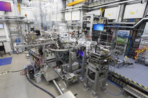
The REMI microscope at the SQS instrument which was used to carry out the current study. Copyright: European XFEL
REMI experimental station at SQS

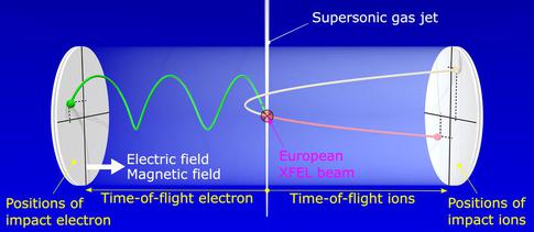
Scheme of a reaction microscope. The molecules are delivered into the interaction region in a supersonic gas jet. The X-ray pulse from the European XFEL hits one of the molecules, creating multiple ions and electrons. Those are guided towards two time- and position-sensitive detectors on opposite sides of a spectrometer by static electric and magnetic fields. In this way, 3D momenta of all recorded particles can be obtained, making it possible to reconstruct the molecular structure by momentum conservation. Copyright: Jahnke, Goethe University Frankfurt
Original Paper:
Gregor Kastirke et al. "Photoelectron Diffraction Imaging of a Molecular Breakup Using an X-Ray Free-Electron Laser" Phys. Rev. X 10, 021052 DOI: https://doi.org/10.1103/PhysRevX.10.021052
